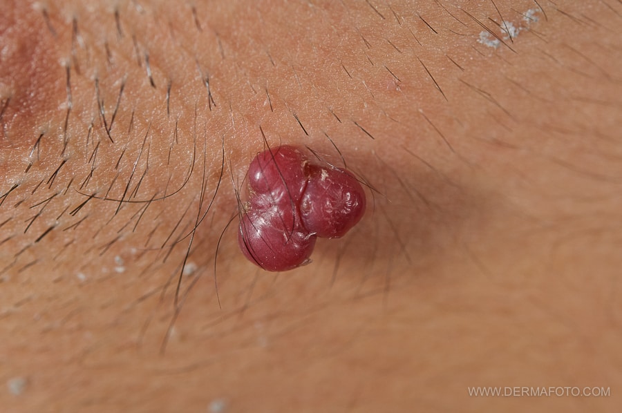
What are ruby points?
Ruby spots or senile angiomas are normal dilations of skin venules. They are part of the normal changes of the skin over time and have no pathological significance: they are benign lesions that are only an aesthetic problem (or discomfort in case they bleed or rub against clothing).
How do ruby spots manifest themselves?
They appear around the third and fourth decades of life, in the form of spots or small cupuliform papules of bright red or dark violet color, with a shiny and smooth surface (hence their name). They are usually asymptomatic, although occasionally they can bleed from rubbing, they can thromboses (they turn black and cause pain for a few days) or they can cause itching or pain when they settle in an area of friction.
Why do ruby dots appear?
There is no specific reason why they appear, although it is common for there to be a genetic predisposition and for them to occur as time progresses (they are part of normal skin aging).
How are they diagnosed?
Usually the trained dermatologist can make the diagnosis with the simple observation of the lesions. However, dermoscopy (use of a magnifying glass with polarized light) can be very useful in cases of doubt, since homogeneous red/purplish vascular lakes are observed inside the ruby spots. Finally, if the diagnosis is still impossible, a biopsy of the lesion can be carried out and observed under a microscope.
Are ruby spots or can they turn into skin cancer?
No. They are benign lesions that have no potential to cause malignant lesions. They do not pose any health problems.
Are they contagious?
No. Ruby spots are simple dilations of the superficial vessels of the skin, they are not the product of any infection, so they are not passed on to other people or on the same patient even if they scratch or touch them.
Are ruby spots very frequent?
Practically all people have ruby spots. They are mainly located on the trunk, although they can appear on the limbs and on the head. They are more common at an older age, and have the same incidence in men as in women.
Do ruby points need to be removed?
It is not strictly necessary to do any treatment, but they can be removed when they pose an aesthetic or practical problem, due to their situation or because they bleed.
How are ruby points treated?
In the past, only electrocoagulation of ruby points could be performed. Although it is a very effective method, it has the disadvantage of requiring local anesthesia (injected into the area to be treated) and that the electrocoagulator is a non-specific destructive procedure: it burns the skin where its tip is applied. Although it is very rare for scars to be left with this method, there is a greater probability than using other much more specific methods, such as laser or pulsed light. Without local anesthesia, cauterization with an electrocoagulator is particularly painful.
Another therapeutic option is to practice cryotherapy with liquid nitrogen and shaving the lesions with a scalpel. This procedure is very fast and is well tolerated in terms of pain. It does not require local anesthesia, since the cold itself reduces the sensitivity of the area. After shaving, it is necessary to use a hemostatic method so that it does not bleed (for example, coagulation with Monsel’s solution or trichloroacetic acid). The risk of scarring is minimal, although if there are very abundant lesions or in noble areas, this technique can be uncomfortable for the patient.
The most comfortable option for the patient and with the lowest risk of scarring is removal using laser or pulsed light devices. The advantage offered by these systems is that they have a specific impact on the haemoglobin that travels within the capillaries that make up the ruby point ( pulsed dye laser, 1,064 nm Nd:YAG laser or pulsed light), or they destroy the redundant tissue of the senile angioma ( CO2 laser) in a very precise way. Any of these systems is comfortable for the patient, can be performed with anaesthetic cream (or without it with the mastery of the parameters of the device) and allows a large number of lesions or bulky angiomas to be treated in a resolute way. In comparative studies, the results obtained with pulsed dye laser and filtered pulsed light are very similar when treating vascular lesions.
In the author’s opinion, in the case of single or small ruby spots, in areas of thick skin such as the back, it is possible to remove them by electrocoagulation or cryotherapy with shaving; but in noble areas, in numerous lesions or when the risk of scarring is to be minimized, treatment with pulsed light and/or laser is very comfortable and satisfactory.
REFERENCES
· Srinivas CR, Kumaresan M. Lasers for vascular lesions: standard guidelines of care. Indian J Dermatol Venereol Leprol. 2011 May-Jun; 77(3):349-68.