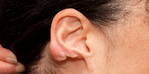
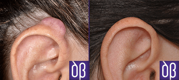
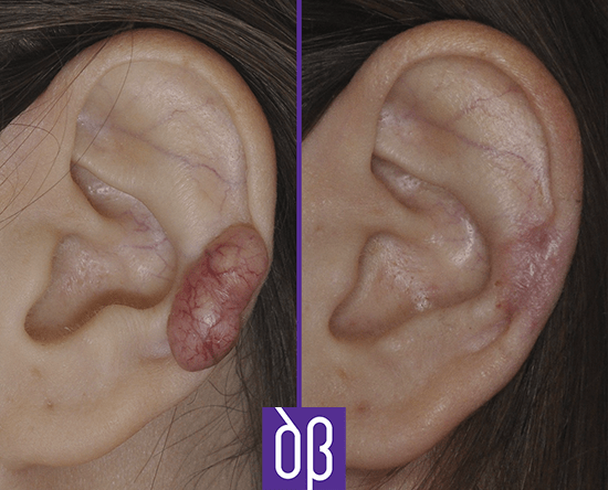
The ear is a structure prone to the formation of keloids, among other reasons because it is an area where incised trauma is usually performed for aesthetic purposes (earrings, piercings, otoplasties). The keloids of the pinna have certain particularities in their therapeutic approach, the main one being that surgical treatment is usually in the foreground. This approach in the management of keloids is very unusual in any other location of the body.
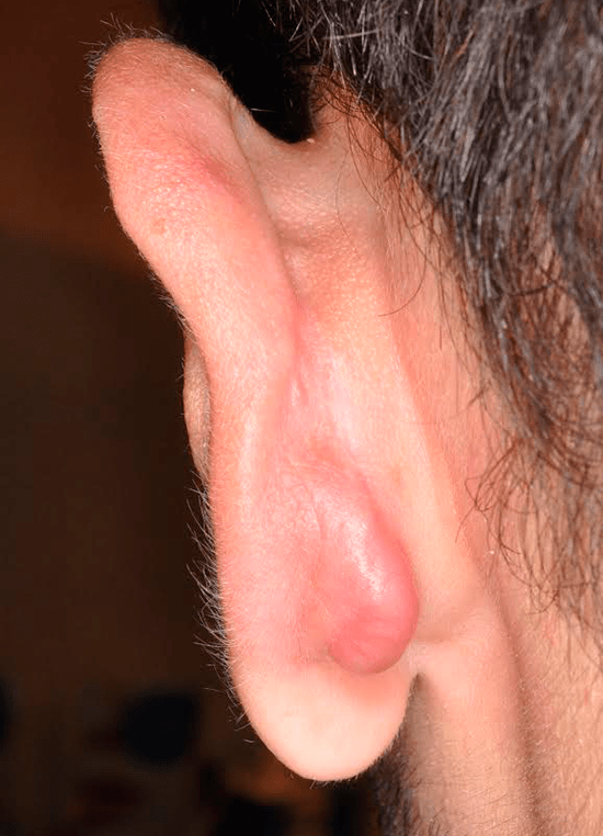
What is the best treatment for keloid of the ear?
There is no therapeutic guide that indicates a specific management direction, and numerous methods are available to treat them. Most authors propose the combination of different therapeutic tools, although in the area of the pinna (unlike the rest of the body), the surgical approach (removal) is usually one of the protagonists. Below we discuss the different therapeutic options that are proposed:
· Silicone gel or patch. The application of silicone products that exert some pressure on the scar is the initial preventive treatment due to its simplicity. This does not mean that it is not effective, as efficacy of 70 – 80% has been reported to prevent the recurrence of hypertrophic scars and keloids.
· Intralesional injection of corticosteroids. Corticosteroids improve keloids through different anti-inflammatory mechanisms, such as reducing glycosaminoglycan synthesis, fibroblast activity and collagen formation. If the keloid in the ear is not very large, they can be used as a first therapeutic option due to their simplicity of administration and speed of action, with favourable responses in 50 – 100% of cases. Although they are a simple and safe option, they are related to some adverse effects such as pain at the time of injection, hypo/hyperpigmentation, the appearance of telangiectasias (capillaries) or atrophy at the injection site in up to 63% of cases.
· Intralesional injection of 5FU. 5-fluorouracil inhibits the proliferation of fibroblasts, the cells responsible for synthesizing collagen. Some authors have reported flattening of keloids in 88% treated with this drug alone. You can learn more about 5FU here.
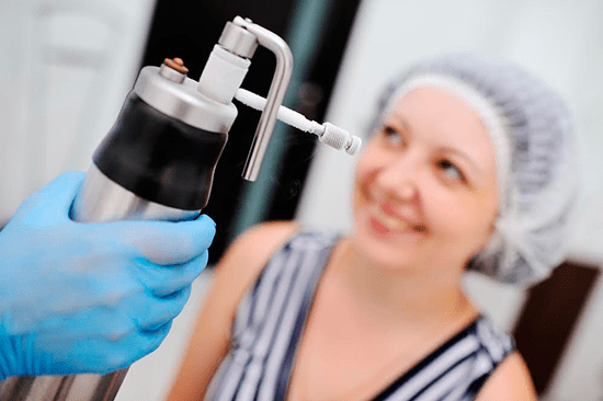
· Cryotherapy. Liquid nitrogen induces by freezing a closure of the vessels that nourish the keloid and, secondarily, a reduction in its size. It is an extraordinarily simple and minimally painful treatment, which is why it is one of the main pillars in keloid therapy. It can be combined with other techniques, such as intralesional injection or lasers. A session can be performed every 3-4 weeks. It is possible that after cryotherapy, blisters or thin crusts may form on the surface of the keloid. Some studies show results of flattening the keloid of up to 73%.
· Surgical excision. The removal of the keloid by scalpel and suture of the skin is usually relatively simple, although it is related to a recurrence rate of 50 – 100%. For this reason, it is exceptional that it is used as the only therapeutic option without combining it with others (radiotherapy, intralesional injection of corticosteroids, silicone occlusive dressings, immunotherapy). There are retrospective studies in which it is observed that the combination of surgery plus infiltration with corticosteroids and occlusive dressings, or surgery plus radiotherapy and intralesional corticosteroids together are superior options to surgery and only infiltration with corticosteroids.
There are studies that combine the excision of the keloid center (“keloid core excision“) or shaving it with subsequent application of intralesional corticosteroids, imiquimod 5%, cryotherapy or silicone patches with excellent results. By means of the “core excision” technique, only the keloid center is removed to drastically reduce its volume and the edges are sutured directly, ensuring that there is no tension in the scar itself.
· Lasers. Several types of lasers have shown efficacy in the treatment of ear keloids, such as CO2, argon, Nd:YAG (more information here) and even pulsed light. Keloid excision with CO2 laser is an alternative to classic surgery and has the advantage that intraoperative bleeding is less – the laser cuts and coagulates simultaneously – and the execution of the procedure is faster than with scalpel and suturing. It seems that an excellent combination of treatments in terms of results is keloid excision, more intralesional application of preventive corticosteroids and post-surgical use of silicone patches (triple therapy).
· Radiation therapy. It is rarely used as a single treatment. It is usually used after surgical removal of the keloid. In these cases, some studies confirm recurrences of less than 10% of cases. Although radiotherapy is generally associated with significant adverse effects, in cases where it is used at an adequate dose and with protection of the surrounding organs, the risk of carcinogenesis is very low.
CONCLUSIONS
· Unlike other areas of the body, ear keloids can be treated by excision, with subsequent application of some additional technique (radiotherapy, intralesional injection of corticosteroids and/or silicone patches).
· Surgical or CO2 laser excision is not recommended as isolated techniques, as the recurrence rate is very high. They need to be combined with other therapeutic options.
· Cryotherapy and intralesional injection of corticosteroids and/or 5-fluorouracil are simple techniques that can be used to reduce the size of keloids in the ear.
REFERENCES
· Sobec R, Dobreanu C, Fodor L, Şomcutean A, Ţichil I, Cosgarea M. Ear keloids: a review and update of treatment options. Clujul Med. 2013; 86(4):313-7.
· De Sousa RF, Chakravarty B, Sharma A, Parwaz MA, Malik A. Efficacy of triple therapy in auricular keloids. J Cutan Aesthet Surg. 2014 Apr; 7(2):98-102.