
What is ringworm? What about onychomycosis?
Nail ringworm is an infection of the nails caused by fungi of the “dermatophyte” type, whether they are the first ones of the hands and/or feet. The term “onychomycosis” simply indicates a fungal infection of the nails and is therefore less specific since it does not indicate which type of fungus is the cause (molds, dermatophytes or Candida sp).
Are nail fungus very common?
Yes. They are the most common cause of nail disorder in adults, accounting for 15 – 40% of all nail diseases. Some studies reveal that up to 3% of adults suffer from a fungal infection (and in people over 70 years of age, up to 50%). The fungi that most frequently infect nails are dermatophytes (the most common is Trichophyton rubrum), which encompass up to 90% of the microorganisms involved.
What promotes nail fungus?
Humidity and heat promote fungal proliferation, as do occlusive footwear, minor repetitive trauma, the patient’s genetic predisposition, advanced age and some concomitant diseases such as diabetes, chronic venous insufficiency or immunosuppression. In up to a third of cases, nail tinya is accompanied by ringworm of the foot (athlete’s foot).
Do nail fungus need to be treated?
Although the main problem of onychomycosis is aesthetic, it is easy for it to cause local discomfort , make it difficult to wear shoes comfortably and also act as a fungal reservoir for future skin infections, so it is recommended to treat fungal nail infections.
What types of onychomycosis are there?
Depending on the pathogenic microorganism, fungal nail infections can take different forms:
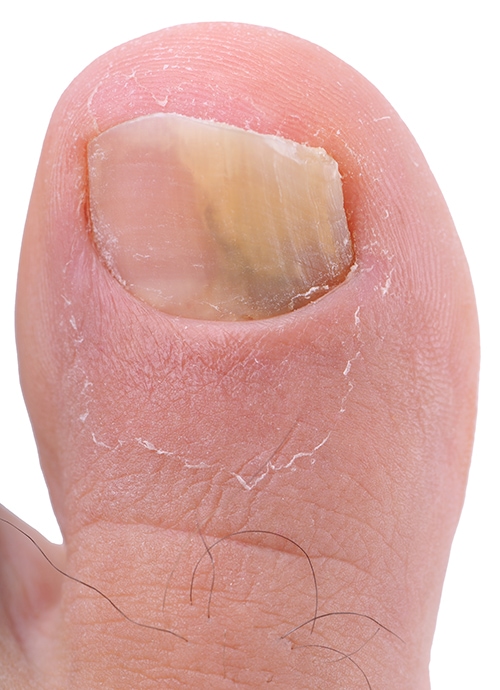
· Distal and lateral subungual onychomycosis. It is the most common form of nail infection, either by dermatophytes (the most common, T. rubrum) or non-dermatophytes. It is more common on the toenails than on the fingernails. The nail thickens and takes on a whitish, yellowish or brown coloration, with a variable degree of separation from the nail bed. It usually affects one nail (the first and fifth toes are the most vulnerable in this regard) and only one foot.
· Proximal subungual onychomycosis. It originates in the proximal fold of the nail and the distal part remains healthy until the condition is advanced. The most frequently involved microorganism is also T. rubrum. It is the least common form in the general population, although it is not uncommon in people infected with the acquired human immunodeficiency virus (HIV).
· Superficial white onychomycosis. The infection begins on the nail surface and progressively spreads and penetrates. Its appearance is that of a white spot that flakes when scratched. This form is more typical of children.
· Endonyx onychomycosis. The fungus infects the nail originally from the center of the nail plate rather than from the margin. There is no separation of the nail from the nail bed and no subungual hyperkeratosis . The blade looks white. The microorganisms most frequently causing this form are T. soudanense and T. violaceum.
· Total dystrophic onychomycosis. Any of the above forms can evolve into this one, where the entire nail plate is destroyed. This variant is usually typical of immunosuppressed patients and the fungus involved is usually Candida sp..
Finally, there are mixed forms of those mentioned above.
How are fungal nail infections diagnosed?
Although the clinical appearance suggests the diagnosis in most cases, it is advisable to proceed with mycological culture of a nail sample. Candida sp. infections have a certain tendency to affect the periungual area (paronychia), although this sign is not pathognomonic.
Microbiological culture is useful as it allows:
– rule out a fungal infection in non-infectious nail disorders
– detect mixed infections
– detect microorganisms resistant to different therapeutic options
What is the treatment for fungal nail infections?
There are topical (locally applied) or oral treatments on the market. Topical drugs are of very little use, since their concentration can drop up to 1,000 times from the nail surface to the bed – which is where it should act – and also because the nail is hydrophilic (attracts water and repels fats) and antifungal drugs are lipophilic (they dissolve in fats). Therefore, topical treatment should only be used in case of contraindication to oral therapy or, in superficial white onychomycosis or in nail involvements of less than 80% of its surface. Topical drugs for the treatment of onychomycosis are recommended only with an evidence level of 3 and a recommendation strength of D (see taxonomic scales here). They include the lacquers of amorolfine, ciclopirox and thioconazole mainly.
The standard treatment of fungal nail infections recommended in European guidelines is oral. The two oral drugs approved for this indication are terbinafine and itraconazole (more rarely fluconazole and griseofulvin can be used).
· Terbinafine. Recommendation strength A, evidence level 1+. It inhibits the formation of the fungal cell wall, and has fungistatic and fungicidal action simultaneously. Its absorption is not affected by food and is eliminated in the urine. Its effect lasts up to 6 months after the end of treatment, as it has a very high half-life. It is very effective against dermatophytes (especially T. rubrum and T. mentagrophytes) but is less effective against Candida than itraconazole. It is a very well tolerated drug, and the most frequent adverse effect is diarrhea, nausea and vomiting, present in only 4.9% of patients who take it. Studies reveal that it is very rare to be associated with hepatic oxidity, so its safety profile is very high. Liver monitoring is only recommended in patients with previous liver disease or in children. It usually has few drug interactions.
· Itraconazole. Recommendation strength A, evidence level 1+. It is active against dermatophytes, yeasts and some moulds. It is not as effective as terbinafine against dermatophytes. Usually, at the doses used in clinical practice, its mechanism is more fungistatic than fungicidal. It is recommended to take it with food, as it increases its absorption. It can be used intermittently (a few tablets taken only a few weeks of the month), as its half-life in the nail is very high. Despite being a very well tolerated medicine, the most frequent adverse effects are headache and gastrointestinal discomfort. Regular laboratory check-ups are recommended, as treatments of more than 4 weeks (e.g., to treat nail infections) in continuous dosage may be related to mild asymptomatic hepatitis in more than 3% of cases. It is contraindicated in patients with heart failure, as it has a negative inotropic effect.
Which treatment is most appropriate and how long should it be taken?
The European guidelines of 2014 recommend terbinafine as the first option for the treatment of dermatophyte nail infections at a dose of 250 mg/day. For nail infections of the hands, therapy should last 6 weeks; and for those of the feet between 3 and 4 months. Itraconazole is administered at a dose of 200 mg/day for 3 months or at 400 mg/day one week per month (one pulse for nail infections of the hands and three for those of the feet). The negative mycological culture rates at 4 months are higher with terbinafine compared to itraconazole, the opposite of the relapse rate after the end of treatment. This, added to the lower risk of drug interactions from terbinafine, makes it recommended as a first therapeutic option. Terbinafine pulses have shown conflicting efficacy data and a regimen that is not continuous is not currently recommended.
For Candida sp. nail infections, itraconazole offers superior results than terbinafine and is the first choice.
Nail infections caused by molds (Aspergillus sp., Scopulariopsis sp., Fusarium sp., Acremonium sp.) are difficult to eradicate and it is not uncommon for nail avulsion or part of it to be required in addition to oral treatment.
How effective is laser treatment of fungal nail infections?
Infrared diode laser devices of 870 and 930 nm have been successfully used, as well as neodymium:YAG of 1064 nm. However, the few studies regarding these treatments do not allow conclusions to be drawn either about their efficacy in the general population or in terms of the duration of their effect.
REFERENCES
Ameen M, Lear JT, Madan V, Mohd Mustapa MF, Richardson M. British Association of Dermatologists’ guidelines for the management of onychomycosis 2014. Br J Dermatol. 2014 Nov; 171(5):937-58.