What are keloids?
They are abnormally large scars, which extend beyond the area of the wound where they originated. Wound repair tissue is formed by collagen fibers: if these proliferate excessively, they can form hypertrophic and keloid scars.
How do they manifest themselves?
They are pink or skin-colored scars, protruding from the edges of the wound with a smooth and shiny surface, and with an indurated touch. They characteristically appear late after the wound.
Why do keloids come out?
They are the product of an alteration in the formation of collagen fibers in the healing tissue. The tendency to form keloids depends on the subject’s own idiosyncrasies , their race, the area of the body where the wound occurs, the type of injury that causes the scar and the care of the wound during healing. Although most of the time they appear as a result of trauma or a wound, their spontaneous appearance has been described for no apparent reason.
Do all people form keloids?
The vast majority of people, depending on the factors mentioned above, are at risk of forming keloids. However, they are more common in black people, in some areas (chest, upper back, shoulders, abdomen, earlobes and joints) or after certain types of wounds (burns, abrasions, dirty wounds).
Where can keloids appear?
Practically in any area of the body, although the most frequent keloids are those secondary to cardiovascular surgery (sternum), abdominal surgery (cesarean section, appendicitis), acne (back, shoulders, chest) and piercings (navel, ear). Interestingly, the face is an area where they rarely appear.
Are keloids dangerous?
No, the vast majority of the time they are an exclusively aesthetic problem, although it is not uncommon for the subject to perceive itching, pain, tightness or pinpricks in the affected area. Sometimes they can be extremely painful to palpation.
How are keloids diagnosed?
In general, the dermatologist makes the diagnosis by visual examination. Very rarely, a skin biopsy can be performed when there is diagnostic doubt, to differentiate it from other rarer lesions such as dermatofibrosarcoma protuberans.
How can keloids be avoided?
Preventing the formation of keloids is practically impossible if the conditions are optimal for their formation. There are people and areas that have a tendency to generate an excess of collagen fibers. In recent traumatic or surgical wounds, and in risk body areas, you should try to do prophylaxis with special care during healing. It is advisable to clean the wound and use dressings made of synthetic polymers such as silicone (gel or patches).
By means of these simple measures while the wound is in the process of healing, in most cases an improvement in the final appearance is observed, although the formation of the keloid is not prevented. Massaging the wound during the scar formation process does not have proven effectiveness .
How are keloids treated?
Many therapeutic modalities are available, so there is no single treatment that is definitive or effective for all people. The standard management of these is carried out through the application of silicone occlusive dressings, corticosteroids (topical or injected into the lesion), cryotherapy (liquid nitrogen), the local injection of antiproliferative drugs (they slow down cell replication), laser devices or a combination of them. Depending on the type and number of keloids, their location and the discomfort they cause, one or another treatment modality is chosen, always personalized for each subject.
What is the most effective treatment for keloids?
There is no single treatment that is clearly better than the others. Contrasting studies on the treatment of hypertrophic scars and keloids show highly variable results, so several therapies are usually combined to achieve the best possible result. What is agreed is that the earlier the treatment in the healing process, the better responses are obtained. In the opinion of the author of this article, the treatment with the best results is obtained, with an excellent risk/cost/benefit ratio, is the early application of non-ablative fractional laser, with or without corticosteroids and cryotherapy combined.
Do keloids have to be removed with surgery?
In general, no. Although there are studies in which surgical excision combined with the injection of local corticosteroids prevents them from reappearing in more than 50% of cases, it is not a standard therapy. The appearance of keloids indicates that the healing process in that area or in that person is not optimal, so it is necessary to be very cautious to recommend the removal of the keloid. In the case of surgical extraction, it is essential to start treatment early after it, applying laser devices or other therapies from the beginning if necessary.
Can keloids be completely removed?
To date, it is not yet possible. Very important improvements can be achieved, up to 70-80%, although a complete disappearance of the lesion is impossible. The earlier and more consistent the treatment, the better results are obtained.
What is the best time to start treatment, once the wound has been made?
Ideally, you should consult a dermatologist as soon as you perceive that the scar is excessive for the wound causing it, or when it causes discomfort. In optimal conditions, it is best to start the maximum treatment between 3-6 months after the start of healing.
Is laser the best treatment for keloids?
There are several types of lasers, and the studies that have been presented regarding their response are highly variable. It is now generally accepted that a complex keloid usually requires (together or not with other therapies) the use of a laser device to optimize the response. The laser is one more therapeutic option that, due to its safety profile, efficacy and few adverse effects, is considered very valid for the treatment of most keloids.
What types of lasers are the most effective for treating keloids?
There are different types of laser devices for the treatment of keloids. The most classic options include the pulsed dye laser (combined or not with corticosteroids, 5 fluorouracil or intralesional bleomycin) and resurfacing with CO2 ablative laser or Erbium:YAG. These two options have been shown to be effective, although it is relatively common for them to have adverse effects for weeks (purpura (reddish spots), erythema, crusts or depigmentation. In recent years, a new type of laser device has been applied, the 1,540 nm fractional non-ablative laser, which not only improves keloids but also hypertrophic, atrophic and hypopigmented scars. The only adverse effect observed with this type of laser is redness and swelling of the treated area, which usually disappear within a few hours. In a comparative study between pulsed dye laser and non-ablative fractional laser, up to 83% of patients preferred the latter.
The opinion of the author of this article is that the 1,540 nm fractional laser has a very high safety and comfort profile, with an excellent response rate with improvements of up to 80% in terms of thickness, keloid area and coloration. The cost/benefit ratio is also optimal:
Keloid by cesarean section treated with non-ablative fractional laser
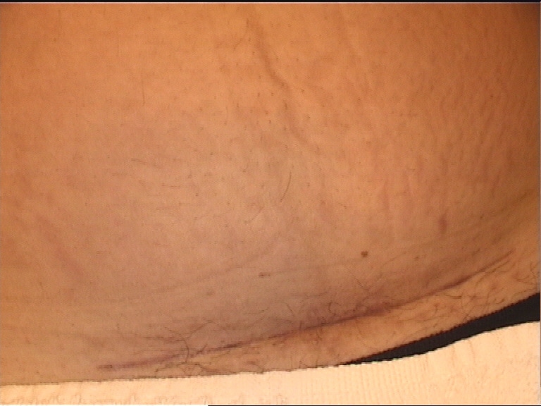
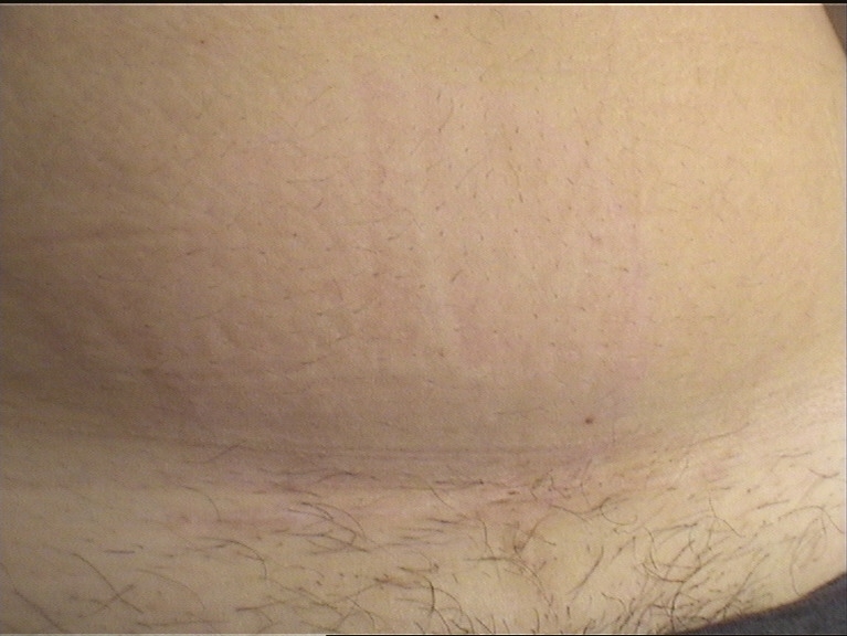


How does the 1540 nm non-ablative fractional laser work for the treatment of keloids?
The fractional laser forms coagulated microscopic columns in the deep tissue of the skin (dermis) without injuring the most superficial layers of the skin (epidermis). These columns of treated tissue are the basis for the formation of new collagen, this time in an organized way, which will replace the old aberrant fibers. Ideally, treatment should begin in the first months after healing (or during it). The sessions, which usually last a few minutes, are carried out 4-6 weeks apart. The number of sessions varies depending on the patient, the number and type of keloids, and the response obtained, although usually between 4 and 6 are needed.
VIDEO ON THE TREATMENT OF SCARS AND KELOIDS
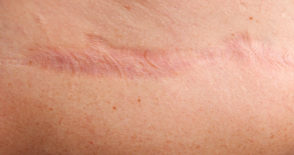
Click here for more information on laser treatment of scars and keloids.
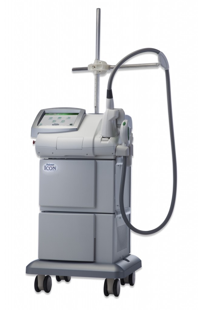
Click here to learn more about laser treatment of acne scars.
References
• Gupta S, Sharma VK. Standard guidelines of care: Keloids and hypertrophic scars. Indian J Dermatol Venereol Leprol. 2011 Jan-Feb; 77(1):94-100.
• Khatri KA, Mahoney DL, McCartney MJ. Laser scar review: A review. J Cosmet Laser Ther. 2011 Apr; 13(2):54-62.