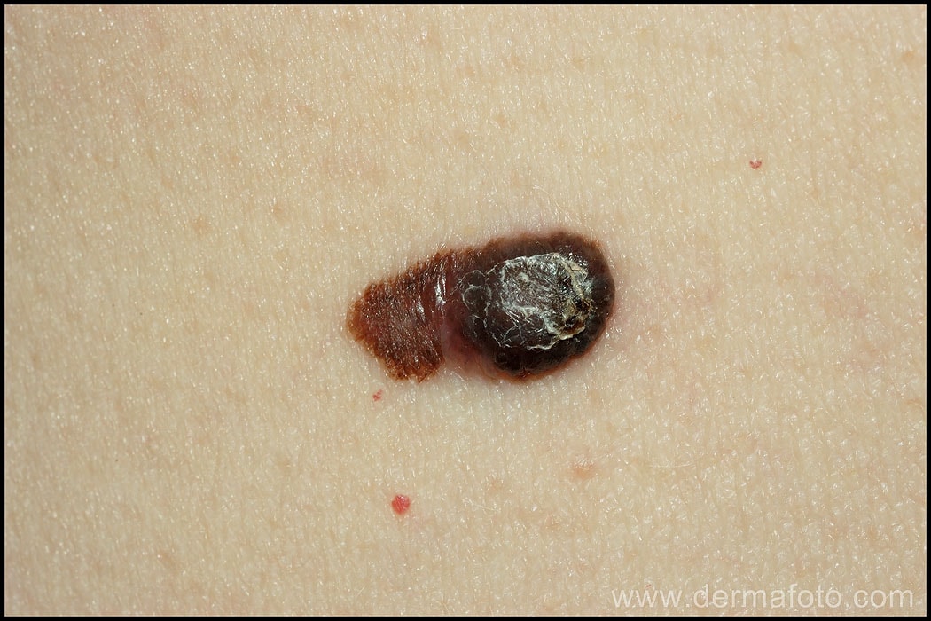
What is melanoma?
It is cancer derived from melanocytes, the cells that give skin its colour. It is a tumour with aggressive behaviour, which has a rapid tendency to grow locally and spread to other organs (metastasis), with a significant resulting mortality if it is not diagnosed early.
Is it a very common tumour?
Fortunately, statistically it accounts for only 4% of skin tumours, but it is the main cause of deaths from skin cancer (6 out of 7 deaths for this reason are caused by this type of cancer).
Its incidence increases annually by 3-7%, especially in young people; and its mortality in Spain has also risen in the last 70 years.
Why does it appear?
The main risk factor for melanoma is a history of sunburn during childhood and youth (more than doubling its probability) and intermittent sun exposure (almost doubling its probability).
Ultraviolet radiation from the sun or tanning booths causes errors in the genetic material of melanocytes (mutations) that, when they reach a limit, can cause the uncontrolled proliferation of abnormal cells.
In addition to the sun habit, there are other risk factors that increase the probability of suffering from this tumor:
· Phototype. It is about the skin’s tendency to sunburn and its ability to tan. It is genetically determined. Light phototypes that burn easily with sun exposure (I and II) have a 3 times higher risk than dark phototypes of melanoma.
· Hair and eye color. People who are blonde, red-haired, light-eyed, and prone to ephelides (“freckles”) have a higher risk of melanoma.
· Presence of moles. Having a high number of melanocytic nevi (moles and freckles) is a risk factor for this type of cancer, especially if these nevi are dysplastic (atypical) – in this case the risk can be multiplied by up to ten.
· Family history of cancer. A family history of melanoma, pancreatic or breast cancer increases the risk.
· Personal history of melanoma. The probability of having a second is 3-6%.
What does it look like?
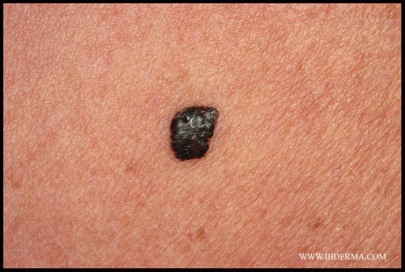
Although there are four types and each one has its own particularities, we can accept that the appearance of melanoma is that of a macula (spot) or papule (raised lesion) of brown-black-reddish coloration, irregular and progressive growth . The most common clinical signs are black coloration and growth.
Where can it appear?
It can appear in any location where melanocytes exist. This implies that it will mainly be on the skin, although it can also be located on the oral or genital mucous membranes , nails, eye, gastrointestinal tract or leptomeninges (central nervous system).
What types are there?
There are four main types:
· Superficial extension. It is the most frequent (70%). It usually appears in the 4th and 5th decade of life in areas intermittently exposed to the sun, such as the back in men and the legs in women. In half of the cases it forms on an old mole, the remaining half appear on healthy skin. Its appearance is that of an irregular, brownish-black spot, of progressive growth. In its development, it initially expands radially for an indefinite time of months or years, and later begins the vertical growth phase, which will determine its invasive power and prognosis. If it is diagnosed in the radial growth phase and only affects the highest layer of the skin (epidermis), it is melanoma in situ, which lacks invasive capacity and whose prognosis is excellent.
· Nodular. Its frequency is 15-20%, and it is more common in males and in the trunk. It manifests as a fast-growing papule (protruding lesion), which can be black or red (amelanotic melanoma). The nodular variety is the one with the worst prognosis, since it does not have a radial growth phase but develops vertically from its beginning. It is not uncommon for it to ulcerate and bleed.
· On malignant lentigo (Dubreuilh’s melanosis). Frequency of 5-10%. It is the typical form of the elderly. It is usually located in areas of chronic sun exposure (characteristically on the face), manifesting itself as a blackish-brown spot with very slow radial growth over years or decades – lentigo maligna or melanoma in situ. After this phase, invasive melanoma forms on the lentigo maligna.
· Acral lentiginous. It corresponds to 10% of this type of skin cancer in the white race, but it is the most common in the black and Asian race. It is located on the palms or soles, manifesting as an ill-defined brown or black spot that progressively spreads until it forms nodules. It is possible that they are located in the bed and/or nail matrix.
What can be done to reduce the risk?
The most important measure is, without a doubt , protection against natural ultraviolet radiation (Sun) and artificial ultraviolet radiation (UVA tanning booths). This is done through the use of chemical and physical sunscreens and with hygienic measures in terms of sun habit. You will find detailed information here.
It is essential to carry out periodic check-ups with Dermatology of moles and freckles, and to consult preferentially when a skin lesion appears with alarming signs (click here for more information).
How is it diagnosed?
The fetal diagnosis is made by excision of the lesion and its study under an optical microscope. The biopsy of a lesion suspicious of this type of cancer must be carried out completely whenever the location allows it: this involves removing the entire lesion and not just a small sample of it. In this way, the thickness of the lesion can be studied, which will indicate its prognosis and treatment.
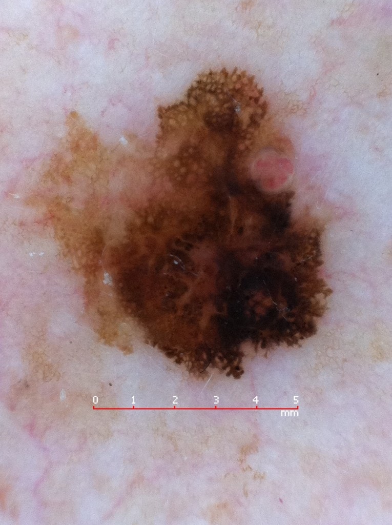
However, the experienced dermatologist can often suspect or diagnose it by means of dermoscopy. This test consists of observing the lesion using a magnifying lens with polarized light, which allows us to distinguish suggestive or defining signs of melanoma in a completely innocuous way. Although dermoscopy allows it to be diagnosed, the excision of the lesion is absolutely mandatory to study its thickness, since it is this that determines the therapeutic conduct.
What diagnostic tests are ordered?
In the first place, it is strictly necessary to have it completely removed and histologically studied under a microscope, as it will dictate its therapeutic conduct and prognosis.
Your dermatologist will then assess whether it is necessary to order any additional complementary tests, such as the study of the lymph nodes in the area, an abdominal ultrasound, a chest X-ray, a blood test or any other test that is considered appropriate to complete the study of the specific case. Diagnostic tests are usually routinely ordered as a screening mode, to check if the disease is localized to the skin or if it has spread to other distant tissues.
How is it treated?
Its treatment depends on the thickness of the lesion (Breslow’s index, indicated in millimeters), as this is directly related to the invasive capacity of the tumour. For this reason, complete excision of the lesion is an essential part of the diagnosis and constitutes the first stage of treatment.
Once the specimen has been extracted, it is observed under a microscope, its thickness is measured (Breslow and Clark indices) and other secondary prognostic criteria are determined: cell replication index (mitosis), invasion of nerve or vascular structures, presence of ulceration or regression, etc.
Depending on the Breslow index and the characteristics discussed, the surgical safety margins around the removed lesion are widened to reduce the probability of local relapse. The amplitude of these margins depends on the Breslow index of the lesion.
Likewise, if the melanoma is of sufficient thickness, the sentinel node should also be studied. This is the first station where a hypothetical spread would take place, as it collects all the drainage from the area of skin where the primary tumour was located. The study of the sentinel node allows us to know if the tumour under study is only a local disease or has caused some metastasis in the lymphatic system. If it is positive, it is necessary to proceed to a complete lymph node emptying of the territory.
In cases of tumours with a Breslow index greater than 4 mm or resected lymph node involvement, interferon alfa, a type of immunological therapy that attempts to form antibodies against tumour cells, is usually used.
In essence, the classic therapeutic scheme for melanoma is as follows:
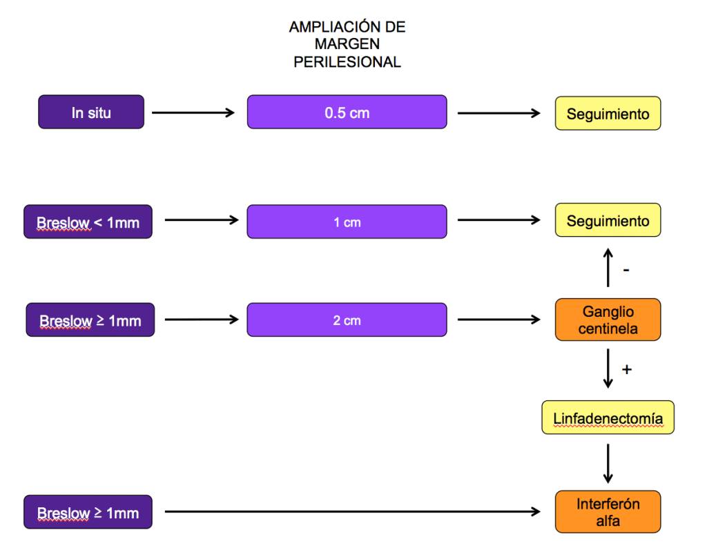
Although this would be a standard diagram, each patient is subjected to a personalized evaluation and the diagnostic-therapeutic maneuvers may vary.
It should also be noted that in the therapy of this type of cancer, more and more advances are being made in immunological and genetic therapy in reference hospitals. Consult your dermatologist for more information.
Who is the doctor who diagnoses and treats it?
Ideally, it should be a dermatologist experienced in dermoscopy and with surgical skills in its removal and, if appropriate, sentinel lymph node. It is common for the treatment of this type of skin cancer to be carried out by a multidisciplinary medical committee that includes different specialists such as dermatologists, oncologists, radiologists, pathologists and/or plastic surgeons. The global approach is always positive for the patient.
REFERENCES
· Guide to the prevention and treatment of melanoma of the Generalitat Valenciana. 2.006.
ACKNOWLEDGEMENTS
To Dr. Leo Barco for the clinical images. Check out his dermatology atlas online.