What is a mole or freckle?
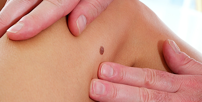
Melanocytic moles, freckles or nevi are benign proliferations of melanocytes, the cells that give color to the skin. It is a normal finding in the skin and consists simply of a grouping (“nevus”) of melanocytes (“melanocytic”), whose appearance derives from a genetic predisposition modulated by more or less intense sun exposure throughout life.
What do they look like and what types of moles are there?
Their appearance varies depending on the type of melanocytic nevus in question, although the most common are classified according to their depth at the junction between the epidermis (superficial layer of the skin) and the dermis (intermediate layer):
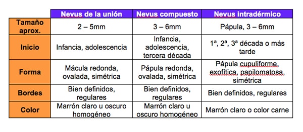
· Melanocytic junction nevus (“freckle”). These are the most common and superficial, and consist of a benign melanocytic proliferation that is located in the basal layer of the epidermis, in contact with the dermis. Clinically, they are brown macules (flat spots), usually rounded in shape and with a size that does not usually exceed one centimeter. Dermoscopically, they are characterized by a typical peripheral pigmented reticulum.
· Compound melanocytic nevus. They have a part that is at the dermo-epidermal junction and in the papillary dermis (upper part of the dermis, the middle layer of the skin). It is usually a lesion with a brown macular component and a raised area (papule) in the center of it, usually of a lighter color.
· Intradermal melanocytic (“mole”) nevus. Intradermal melanocytic nevi are deeper and nest in the reticular dermis and subcutaneous cellular tissue (skin fat). They manifest as exophytic (very protruding) brown papules, which is why they are popularly called “warts” incorrectly.
Are melanocytic nevi bad or can they be bad?
Melanocytic nevi are an accumulation of normal skin cells and do not have any intrinsic malignancy in themselves. However, when these melanocytes proliferate and adopt irregular morphology (dysplastic melanocytic nevus) they can have a risk of degeneration to melanoma. Dysplastic melanocytic nevi are relatively common in young patients and most of them do not finally transform into melanoma. For this reason, it is important that routine check-ups with dermoscopy are carried out periodically with the dermatologist. In this way, we determine if the freckles or moles are conventional, have a risk of degeneration or it is possible to detect an incipient malignant lesion.
Why do moles and freckles appear?
The appearance of melanocytic nevi is mainly regulated by the genetic component of the person, although sun exposure can favor the appearance of moles and freckles in greater numbers. The use of sunscreen reduces the development of melanocytic nevi in areas exposed intermittently to the sun.
Do moles have to be removed?
Moles and freckles do not necessarily have to be removed: most of the time it is done for aesthetic purposes. Occasionally, if the dermatologist observes that the pigmented lesion is irregular or requires further study, a complete surgical excision and a histological study (biopsy) will be performed.
How do you remove moles?
There are different methods to remove or eliminate moles. The choice of one technique or another will depend on several factors: type of lesion and location, purpose of removal and patient preference. The techniques to be used are the following:
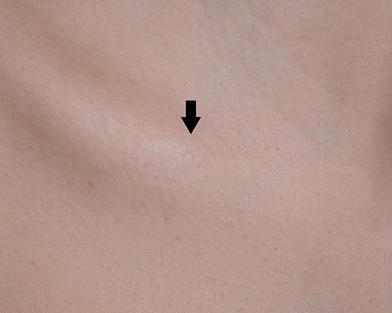
· Spindle surgery. Scalpel excision and thread suturing is performed whenever there is diagnostic doubt about the pigmented lesion: it is the technique of choice as it allows the histological study of the entire piece. It is performed under local anesthesia and after the procedure there is usually a scar that may be more or less visible. Surgery is the optimal option in certain locations (e.g., the nasolabial fold) or if the mole is particularly voluminous. If the purpose of the mole removal is aesthetic, it is possible to choose other options that have a lower risk of scarring (e.g., shaving or CO2 laser).
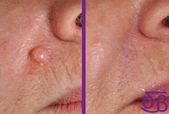
· Surgical shaving. It is performed on exophytic lesions (protrusions). It is performed by local anesthesia or cryotherapy with nitrogen and subsequent tangential slicing with a scalpel. In this way, the superficial part extracted from the tooth can be analyzed histologically, but not the deep portion that remains in the skin. Analogously, it could be compared to cutting a tree with a chainsaw: the trunk and branches are removed, but the root is left on the ground. It is a very fast procedure with a favorable aesthetic result in the vast majority of cases. In some cases, the remaining portion of melanocytic nevus can be repigmented in the months after the procedure and take on the appearance of a completely flat freckle and especially black. This technique has fewer risks and complications than surgery, but the probability that the mole will not be completely removed is higher (between 13 and 20% according to studies).
· Depigmenting lasers. This type of device (pulsed light, Alexandrite) specifically impacts on melanocytes and destroys them. It should only be performed on flat, non-bulky melanocytic nevus. These types of techniques are NOT routinely recommended, as they do not allow the histological study of the removed lesion. Ideally, this system should only be used when there are remains of a previously analysed melanocytic nevi (e.g., after a surgical shave to remove the remaining base). There have been cases in which the superficial part of a melanoma has been accidentally removed by laser (false disappearance), making it impossible to diagnose it early and favouring the undetectable progression of the tumour. Always consult a dermatologist before proceeding with this treatment system.
· Electrocoagulation. Using an electrocoagulator or electric scalpel, it is possible to destroy tissue by heat. This technique is used for intradermal (protruding) melanocytic nevi. The heat damage of the electrocoagulator is poorly controlled by the dermatologist and can cause a scar on the skin, so extreme caution should be exercised in this regard. The destruction of the tissue does not allow histological study (biopsy) either. Nowadays it is more common to opt for a more precise device such as the CO2 laser, which is more safe.
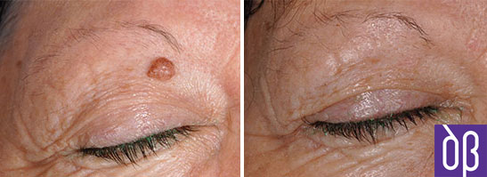
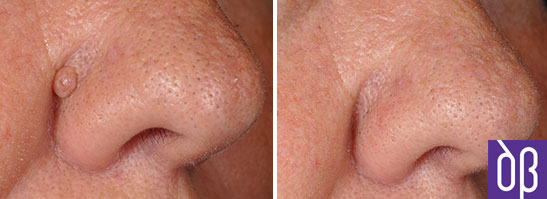
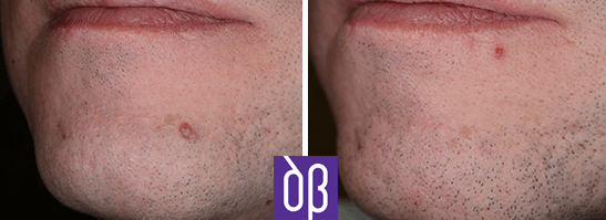
· CO2 laser. The CO2 laser instantly removes tissue where light hits. It can be used as a scalpel to cut (instead of a metal scalpel it would be light) or as a tool to vaporize protruding lesions. Its advantages are its high degree of precision and the control you have over the scope of its depth, so the probability of leaving a scar in the treated area is minimal. Current guidelines recommend that the ultra-pulsed CO2 laser (pulses < lasting 1 millisecond) be used in a first treatment to remove the volume of the mole and, if required, in a second stage weeks later a depigmenting laser (Alexandrite, neodymium:YAG or pulsed light) to remove the residual pigmented part. This therapeutic combination is the one that produces the best aesthetic results and the lowest rate of complications.
ABSTRACT
· You should always consult a dermatologist to assess whether that mole or freckle presents potential signs of malignancy through dermoscopy or biopsy if there are doubts.
· It is recommended, whenever possible, to histologically analyze the removed melanocytic nevus.
· According to current therapeutic recommendations, most melanocytic nevi can be satisfactorily eliminated with the combination of different lasers, this option being the one that offers the best outcome/cost-risk ratio compared to others (surgery or electrocoagulation).
· Even using the laser, it is more than likely that for the complete removal of a mole or freckle more than one laser will have to be used and the treatment must be performed in more than one session because moles have variable depths: this reduces the risk of scarring and ensures the complete removal of the lesion.
· It is normal and relatively common that after a first treatment part of the mole reappears in the form of a flat brown or black spot (remnants of the deep part of the melanocytic nevus). This can be removed by depigmenting or ablative laser.
REFERENCES
· Sardana K, Chakravarty P, Goel K. Optimal management of common acquired melanocytic nevi (moles): current perspectives. Clin Cosmet Investigaig Dermatol. 2014 Mar 19;7:89-103.