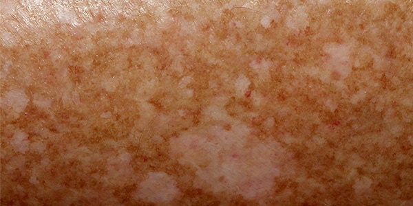
What is idiopathic hypomelanosis in droplets?
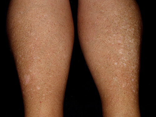
Idiopathic guttate hypomelanosis is a skin pigmentation disorder characterized by the appearance of white, round or oval spots, usually less than one centimeter, on areas of the body chronically exposed to the sun. These spots are especially common on the front of the legs, forearms, upper back, and décolleté.
Is it a very common disorder?
Yes. In sunny countries, the vast majority of the population that has been exposed to the sun, actively and/or passively, has this disorder. It is the most frequent cause of white spots on the legs; and this is especially common in middle-aged women with fair skin. Around 30 years of age it is more common in women. However, with the passage of age, the frequency equalizes between men and women.
What causes idiopathic hypomelanosis in droplets?
It is unknown, although it is known that chronic sun exposure is the main precipitating factor. It is the translation of a localized reduction in the number of melanocytes (the cells that give color to the skin) and melanin (the pigment contained in melanosomes of melanocytes).
What is the implication of idiopathic guttate hypomelanosis?
It is a benign disorder, with no risk of degeneration to skin cancer and no relation to vitiligo. However, as it is a consequence of chronic sun exposure, it indicates that the patient’s skin has received a significant amount of ultraviolet radiation throughout life, so caution must be exercised with skin check-ups since in these cases the incidence of pre-malignant lesions (actinic keratosis) and malignant lesions (basal cell carcinoma, squamous cell carcinoma and/or melanoma) may be more common.
How is hypomelanosis diagnosed?
Usually the diagnosis is clinical (observation). Exploration using Wood light and/or dermoscopy may be useful. Very exceptionally, a skin biopsy will be performed if there are diagnostic doubts that may imply a prognostic difference for the patient.
What is the treatment for guttate hypomelanosis?
There are different options, so there is no one that is clearly superior to others. Topically , corticosteroids, calcineurin inhibitors (pimecrolimus) and/or retinoids (tretinoin, derived from vitamin A) can be used. As for surgical methods, dermabrasion, cryotherapy or skin micrografts can be used. However, since these techniques are associated with discomfort and a subsequent recovery period due to the injury they cause to the skin, interest has focused on the use of lasers. Indeed, the most recent studies reflect the efficacy of the erbium non-ablative fractional laser, with which documented improvements of up to 85% are achieved. The CO2 ablative fractional laser produces similar improvements with fewer sessions, but the appearance of residual crusts and the risk of residual hyperpigmentation recommend caution with this procedure.
NOTE
Thanks to Dermatoweb.net for the excellent clinical photographs.
REFERENCES
· Rerknimitr P, Disphanurat W, Achariyakul M. Topical tacrolimus significantly promotes repigmentation in idiopathic guttate hypomelanosis: a double-blind, randomized, placebo-controlled study. J Eur Acad Dermatol Venereol. 2013 Apr; 27(4):460-4.
· Rerknimitr P, Chitvanich S, Pongprutthipan M, Panchaprateep R, Asawanonda P. Non-ablative fractional photothermolysis in treatment of idiopathic guttate hypomelanosis. J Eur Acad Dermatol Venereol. 2014 Oct 10.