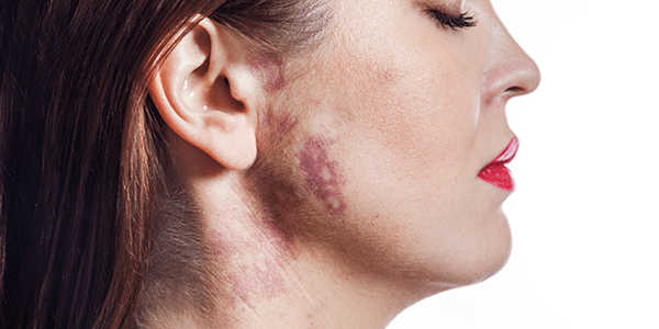
What are vascular lesions and how is the treatment of vascular lesions with laser and pulsed light?
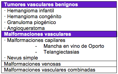
Vascular lesion is understood to be any cutaneous manifestation that originates in a blood vessel, whether normal, aberrant or pathological. Among the most frequent vascular lesions are the following (subtypes are not included):
Certain lasers and filtered intense pulsed light can selectively impact the hemoglobin contained in the blood of the vessels. The impact of this light energy on the vessel causes a collapse in its walls and it closes or attenuates, improving the clinical appearance of the vascular lesion.
Which vascular lesions can be treated with laser or intense pulsed light?
The vascular lesions that are most frequently treated with lighting systems are the following:
· Telangiectasias (“broken capillaries”)
· Rosacea (couperose)
· Spider Veins
· Venous Lake
· Ruby spot (senile angioma)
· Civatte poikiloderma
· Pyogenic granuloma
· Infantile hemangioma
· Malformation of caps (stain in Port Wine)
· Angiofibroma
· Varicules, spider veins, and capillaries of the legs
· Hypertrophic and keloid scars
· Angiokeratomas
Treatment of vascular lesions with laser and pulsed light: what types of lasers are used.
Lasers that specifically impact blood vessels (haemoglobin) or non-specific lasers can be used, which simply destroy skin tissue (CO2 or ablative erbium). The first group includes the KTP laser (532 nm), the pulsed dye laser (585-595 nm) and the Nd:YAG laser (1,064 nm). Pulsed light is not a laser, but if it is technically filtered, it can affect haemoglobin and is also useful for treating vascular lesions.
What is the best laser to specifically treat each vascular lesion?
Below we will discuss, according to the guidelines of the European Society of Dermatology for Laser ( SEDL), the optimal laser treatments for the different vascular lesions, according to their degree of scientific evidence in parentheses. We will not talk about infantile hemangiomas due to their treatment specificity and the advent of oral beta-blockers a few years ago, which changed the conception of therapy for this type of benign tumors.
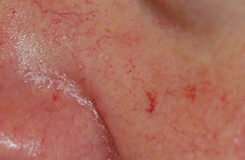
· FACIAL TELANGIECTASIAS AND DIFFUSE ERYTHEMA. They can also be popularly referred to as ‘broken capillaries’ of the face and facial couperosis. Telangiectases or capillaries can be simple (linear), arboriform (spiders), punctate or papular (“pimple”). They are common on the nose, cheeks and chin, and have a diameter of between 0.1 – 1 mm.
Different studies have confirmed the efficacy of pulsed dye, KTP and pulsed light lasers for facial telangiectasias, with clearance responses of 50 – 90% with 1 – 3 sessions. Other lasers such as long-pulse alexandrite or ultra-short-pulse neodymium may incur more risks.
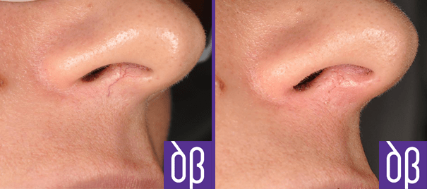
Short-wavelength devices (KTP, pulsed dye, and pulsed light) are recommended for finer telangiectasias or for diffuse redness. For deeper, bluish, or thicker vessels, higher-wavelength devices (e.g., 1.064 nm neodymium) are preferred.
Click here for more information on laser treatment of facial capillaries.
For facial telangiectasias and diffuse erythema, the SEDL recommends treating them with lasers or pulsed light (grade 1-A). As a first option, the use of pulsed dye, KTP or pulsed light (grade 1-A) laser is recommended. If they do not achieve the desired effect, the use of Nd:YAG or diode laser (grade 1-B) can be tried.
Cooling the epidermis during the procedure reduces discomfort and the risk of damaging the epidermis, so it is recommended to apply it during treatment (grade 1-B). Some laser devices have a skin contact head that is continuously cooled. Additionally, a cold ultrasound gel can be applied to the surface of the skin to further decrease the temperature and increase safety.
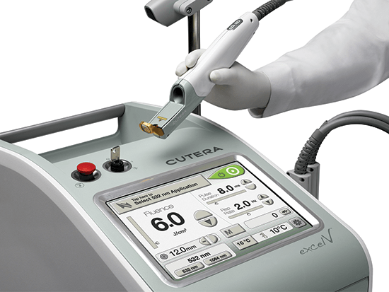
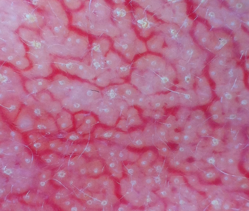
· ROSACEA. It is common for patients with rosacea to report a sensation of flushing (transient redness) or couperose (permanent redness), caused by excessive vasodilation of the dermal capillaries. It is known that this rich vascular structure is related to a progression of inflammatory lesions and disease, so its removal improves the prognosis of rosacea. The only treatment available to eliminate these capillaries is laser or pulsed light, since topical therapies are not effective. Click here for more information on the treatment of rosacea with laser.
Couperose and flushing of rosacea can be effectively and safely treated with pulsed dye laser, KTP, and/or pulsed light (grade 1-A). There is less evidence to support the use of neodymium laser for this indication (grade 1-C).
· ARACNIFORM HEMANGIOMA. It is a thick central telangiectasia from which numerous thinner radial branches start. They are especially common in children, near the nose or eyes. It is not uncommon for more than one session to be needed to remove the lesion completely, since the central vessel is of high flow.
For the treatment of spider haemangiomas, the SEDL recommends the use of millisecond pulse neodymium laser (1,064 nm), KTP laser (532 nm), long pulse pulsed dye laser (585 – 595 nm) or pulsed light with grade 1-B evidence.
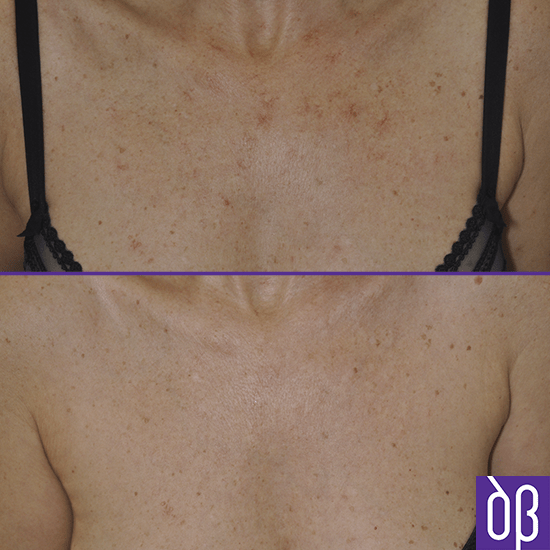
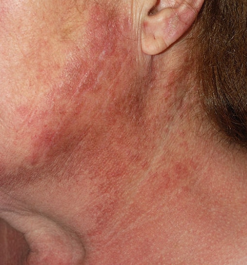
· CIVATE POIKILODERMA. It is caused by chronic sun exposure and is usually very resistant to treatment. It is characterised by the presence of atrophic skin with reddish, whitish and brown spots. It is characteristic in the area of the sides of the neck and on the décolleté. It usually requires several treatment sessions to obtain a noticeable improvement. It is advisable to reduce the laser energy by 20-30% in delicate areas to reduce the risk of burns, avoid overlapping shots and use wide shooting spots (e.g., 10 mm).
For the treatment of Civatte pochyloderma , pulsed light, the KTP laser of 532 nm and the long-pulse pulsed dye laser (evidence grade 2-B) can be used.
· PYOGENIC GRANULOMA. These are benign vascular tumors, usually exophytic, ulcerated and bleeding. They usually appear in children. Although they can be treated with surgical excision, cryotherapy, electrocoagulation, pulsed dye, CO2 and neodymium lasers may be useful (grade 1-C recommendation).
· VENOUS LAKES. These are localized dilations of the venules. They are practically exclusive to the lips. They respond to surgical excision, cryotherapy, pulsed light, pulsed dye laser, neodymium laser and CO2 laser, among other options. The fastest and safest option is the 1,064 nm long-pulse neodymium laser (evidence grade 1-C).
· RUBY SPOT OR SENILE ANGIOMA. It is the most common benign vascular lesion in humans from a certain age. Here you can learn more about its morphology. They can be removed with KTP laser, neodymium, pulsed dye and pulsed light (grade 1-C). In general, the neodymium laser requires only one session for complete removal, although this device can be more annoying than the KTP laser.
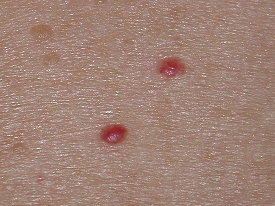
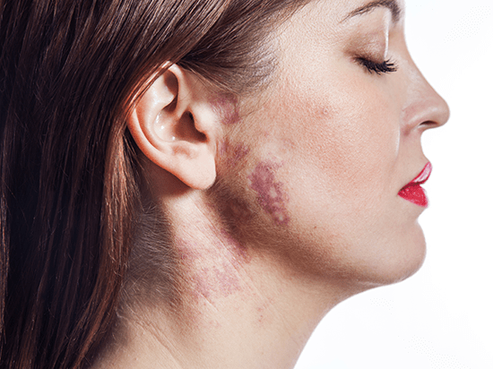
· CAPILLARY MALFORMATIONS (PORT WINE STAIN ). They affect up to 0.5% of newborns. Initially they are flat lesions (macules) and around the age of 30 they can begin to gain volume (hypertrophy). The standard treatment to date has been the long-pulse pulsed dye laser , and it should be borne in mind that up to 20% of port wine stains are resistant to this therapy from the 7th-10th laser session. It is exceptional that they disappear completely even if they have a good response to treatment.
If the treatment of vascular lesions with laser and pulsed light is started before the age of one , it is possible to prevent the progression of the capillary malformation and even make it disappear completely, since the blood vessels are usually more superficial in these conditions. However, although there are authors who recommend early treatment, there are studies that do not support this behavior and confirm a possible neovascularization of the lesion over time once treated. Children over one and a half years old or those who have large capillary malformations require general anesthesia in each session.
The SEDL recommends early treatment of port wine stains, every 2-3 weeks, when pulsed dye laser is used. Numerous sessions of laser therapy are needed, until the capillary malformation stops showing response after each session (grade 1-C).
The degree of response depends on several parameters. Pink lesions tend to respond less than red lesions. Capillary malformations with papules and nodules require longer pulses of energy and longer wavelengths. Port-wine stains smaller than 20 cm2 tend to respond better than larger ones. Capillary malformations of the head and neck (except those of the V2 area of the face) respond better than those of any other location, especially those of the distal extremities.
As a first option for the treatment of capillary malformations (port-wine stain), the SEDL recommends the use of pulsed dye laser, although superficial lesions also respond satisfactorily to KTP laser and pulsed light (grade 1-C).
For those resistant or hypertrophic capillary malformations, it is recommended to use long-pulse neodymium lasers, alexandrite lasers, diode lasers or pulsed light lasers.
Maintenance treatment may be necessary for life, with a laser session every year or every certain period of time. In general, capillary malformations usually lighten by 50% on average. Those stains in port wine associated with Sturge-Weber syndrome or Klippel-Trenaunay syndrome do not respond as satisfactorily as sporadic ones.
· TELANGIECTASIAS, SPIDER VEINS AND VARICULES OF THE LEGS. The effectiveness of light systems is less effective in treating vascular lesions of the legs compared to facial ones. The variability of thickness, flow, depth and type of vessel make these more difficult to treat. The parameter that determines the type of laser to be used is the diameter of the target vessel.
For vessels smaller than 1 mm in diameter, lasers with a wavelength of less than 600 nm (mainly KTP and pulsed dye) can be used effectively and safely. For vessels larger than 1 mm , higher wavelengths (Nd:YAG, alexandrite or diode) and pulses of 1 – 50 ms are recommended. During treatment, it is essential to cool the epidermis to reduce the risk of adverse effects.
Spider veins and leg varicules less than 1 mm in diameter should be treated with KTP or pulsed dye laser (grade 1-A). Alternatively, pulsed light (grade 1-C) may be used. Other types of lasers are secondary for vascular lesions of this diameter.
You can find more information about treating spider veins and varicules of the legs with KTP laser here and neodymium laser here.
When using the neodymium laser, it is advisable to set up spots of 3 – 8 mm, pulses of 30 – 60 ms and fluences of between 250 – 600 J/cm2. Only slight overlapping of pulses with 3 mm spots will be made, working with the 8 mm one is contraindicated.
REFERENCES
· Adamič M, Pavlović MD, Troilius Rubin A, Palmetun-Ekbäck M, Boixeda P. Guidelines of care for vascular lasers and intense pulse light sources from the European Society for Laser Dermatology. J Eur Acad Dermatol Venereol. 2015 Sep; 29(9):1661-78.